When a scan shows a mass on the pancreas, the first worry is often pancreatic cancer. That worry makes sense. But it helps to know one thing early. Not every pancreatic mass is cancer. Some are benign. Some are cysts. Some are areas of inflammation. Some are slow-growing tumors, and some are pancreatic cancer. The only way to know is a careful evaluation.
This article explains what a pancreatic mass is, the warning signs to watch for, who is at higher risk, and the steps doctors take to reach a diagnosis. It also covers how endoscopic ultrasound, often called EUS, helps find and study these lesions when other scans do not give a clear answer.
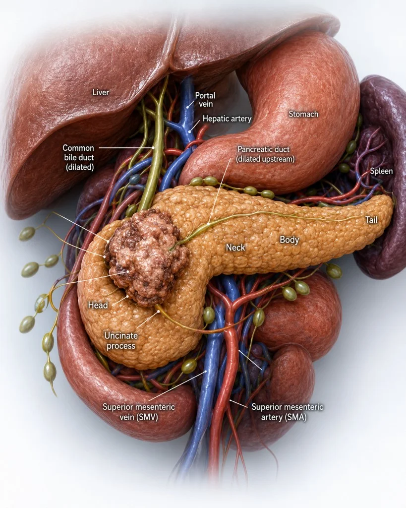
What is a pancreatic mass?
A pancreatic mass is an abnormal spot or growth in the pancreas. In simple terms, it is an area that looks different from the normal gland. It may be solid, or it may be filled with fluid.
The pancreas sits deep in the belly, behind the stomach. Because of this, a mass there often cannot be felt from the outside and may not cause any symptoms in the early stages. That is one reason pancreatic cancer can be hard to catch early.
A pancreatic mass can be many different things. Pancreatic ductal adenocarcinoma is the most common type of pancreatic cancer. But a mass can also be a neuroendocrine tumor, a cystic growth, or swelling from pancreatitis. Some masses look like cancer on a scan and turn out to be harmless. This is exactly why tissue sampling and expert review matter so much.
Solid and cystic lesions
Doctors often start by describing a pancreatic mass as solid or cystic. This simple split says a lot about what the mass might be and how urgent it is.
Common solid masses include:
- Pancreatic ductal adenocarcinoma
- Pancreatic neuroendocrine tumor
- Solid pseudopapillary neoplasm
- Acinar cell carcinoma
- Pancreatic lymphoma
- Cancer that has spread from another organ
- Focal autoimmune pancreatitis
- An inflammatory mass from chronic pancreatitis
Common cystic or mixed lesions include:
- Intraductal papillary mucinous neoplasm, or IPMN
- Mucinous cystic neoplasm
- Serous cystadenoma
- Cystic neuroendocrine tumor
- Pseudocyst
- Walled-off necrosis
Some cystic lesions are harmless and can simply be watched over time. Others carry a risk of turning into cancer and need close follow-up or treatment. Telling these apart is a key part of the workup.
Why the location matters
Where the mass sits in the pancreas changes the symptoms it causes and how it is treated.
A mass in the head of the pancreas can press on the bile duct. This can block the flow of bile and cause jaundice, which is a yellow color in the skin and eyes. It can also lead to dark urine, pale stools, and itching. On imaging, doctors may see both the bile duct and the pancreatic duct widened. This is called the double-duct sign.
A mass in the body or tail can stay quiet longer. It may not cause symptoms until it grows or spreads. When symptoms do show up, they can include belly pain, weight loss, or signs that the mass is involving nearby blood vessels.
A mass in the uncinate process, the hook-shaped lower part of the pancreas, can involve major blood vessels or cause subtle symptoms that are easy to miss.
Warning signs and symptoms
Early pancreatic cancer often has no clear symptoms at all. When symptoms do appear, they can be vague and easy to blame on something else. Signs that deserve attention include:
- Painless jaundice, meaning yellow skin or eyes without any pain
- Weight loss you cannot explain
- Pain in the upper belly that spreads to the back
- New diabetes, or diabetes that suddenly gets worse
- Loss of appetite
- Nausea or feeling full very quickly
- Repeated bouts of pancreatitis
- Greasy, foul-smelling stools or trouble absorbing food
- A widened bile duct or pancreatic duct seen on a scan
- A mass found by chance during imaging done for another reason
Having one of these does not mean you have cancer. Many of these signs come from other, less serious conditions. But they are good reasons to see a doctor and get checked.
Risk factors
Some things raise the risk of pancreatic cancer. Knowing them can help you and your doctor decide when to look closer.
- Cigarette smoking
- Chronic pancreatitis, which is long-term inflammation of the pancreas
- Family history or an inherited syndrome
- Age over 60
- Obesity and diabetes
- Certain pancreatic cysts that can turn into cancer
Not everyone with these risk factors will get pancreatic cancer. And some people who get it have no clear risk factors at all. Still, these help guide who may benefit from closer screening and follow-up.
How a pancreatic mass is diagnosed
Reaching a clear diagnosis usually takes more than one test. It often starts with a scan, moves to more detailed imaging, and, when needed, ends with a tissue sample.
The role of endoscopic ultrasound (EUS)
Endoscopic ultrasound is one of the most useful tools for studying the pancreas. During EUS, a thin, flexible scope with a small ultrasound probe on the tip is passed through the mouth and down into the stomach and small intestine. Because the pancreas sits right behind these organs, the probe can get very close to it and produce clear, detailed pictures.
EUS is helpful in several ways:
- It gives high detail views of small pancreatic lesions
- It shows the size, edges, blood vessel involvement, lymph nodes, and duct changes
- It can find lesions that a CT or MRI missed or could not clearly define
- It allows a tissue sample through a fine needle when that is needed
- It helps check nearby lymph nodes and guide staging
- It can tell solid parts from cystic parts and pick the safest place to sample
When a tissue sample is needed, the doctor can pass a thin needle through the scope and into the mass while watching on ultrasound. This is called fine needle biopsy or fine needle aspiration. The sample then goes to the lab for study under a microscope and, in many cases, molecular testing.
In short, EUS can show tumor size and edges, whether the mass is touching or growing into blood vessels, whether ducts are blocked or widened, the state of nearby lymph nodes, and whether a lesion is solid or cystic. All of this shapes the plan that follows.
The diagnostic pathway
The path from a suspected mass to a treatment plan usually follows clear steps:
1. Suspicion of a problem, or an abnormal scan
2. A pancreas-protocol CT scan, or an MRI with MRCP
3. EUS for a closer look
4. EUS-guided tissue sampling when it is indicated
5. Pathology and molecular analysis of the sample
6. Staging by a team of specialists
7. A treatment plan built for the individual patient
Each step adds a piece of the picture. By the end, the care team knows what the mass is, how far it has spread, and the best way to treat it.
Why early, expert evaluation matters
Pancreatic cancer is often found late because the early signs are so quiet. Finding a mass early, studying it with care, and getting a clear tissue diagnosis can shape the entire treatment plan. EUS plays a central role here because it can reach small lesions that other scans struggle to see, and it can sample them in a safe, targeted way.
Care that brings together gastroenterology, surgery, cancer care, and radiology gives patients the fullest picture and the best chance at the right plan for them.
Access to EUS is not the same everywhere, since it is a specialized test. For patients in Greenville, the Upstate, and across South Carolina, a careful EUS evaluation close to home can help confirm what a pancreatic mass is and guide the next step. You can read more about how endoscopic ultrasound is used to study the pancreas, and about EUS in Greenville, SC and EUS across upstate South Carolina.
Frequently Asked Questions
Is every pancreatic mass cancer?
No. Many pancreatic masses are not cancer. Some are cysts, some are areas of inflammation, and some are slow-growing tumors. A full workup is needed before anyone can say for sure.
Can EUS tell if a mass is cancer?
EUS gives very detailed images and can guide a needle to take a tissue sample. The tissue sample is what confirms whether a mass is cancer. EUS makes taking that sample safer and more accurate.
What is the double-duct sign?
It is when both the bile duct and the pancreatic duct look widened on imaging. It can be a clue that a mass in the head of the pancreas is blocking both ducts at once.
What are the early warning signs of pancreatic cancer?
Some signs to watch for are painless jaundice, unexplained weight loss, upper belly pain that spreads to the back, new or worsening diabetes, and loss of appetite. These can also come from other conditions, so they are a reason to get checked, not a reason to panic.
Who is at higher risk for pancreatic cancer?
People who smoke, have chronic pancreatitis, have a family history or an inherited syndrome, are over age 60, or have obesity and diabetes may be at higher risk. Certain pancreatic cysts can also raise the risk.
Do I need a biopsy for every pancreatic mass?
Not always. The care team decides based on the type of mass, its features, and the overall plan. Some masses are watched over time, some are removed, and some are sampled first to guide the next step.
About the author
Dr. Kalpit Devani is a fellowship trained, board certified gastroenterologist with additional training in interventional endoscopy, based in Greenville, South Carolina. His work focuses on advanced procedures such as endoscopic ultrasound (EUS) and ERCP, which are used to evaluate and manage conditions of the pancreas and bile ducts.
This article is for general education only. It is not medical advice and does not replace care from a qualified health professional. Every case is different, and decisions should be based on a full evaluation by your own care team.
Dr. Kalpit Devani, MD, FACP, FACG, FASGE is a board-certified interventional gastroenterologist based in Greenville, South Carolina, specializing in diagnostic and therapeutic EUS, complex ERCP, and endoscopic suturing.
Related Posts
Post-ERCP Pancreatitis in Malpractice Litigation: A Guide for Defense Attorneys
Post-ERCP pancreatitis is the most common ERCP complication and the most common ERCP malpractice claim. What defense counsel should know about risk, causation, and the record.
Read more →Choosing a Gastroenterology Expert Witness for an Advanced Endoscopy Case
Board certification does not tell you whether a gastroenterologist performs ERCP or EUS. How attorneys should evaluate an expert for an endoscopy case.
Read more →What Are Pancreatic Cysts? When to Worry and When to Watch
Pancreatic cysts are often found by chance on CT or MRI. Learn the main types—including IPMN, serous, mucinous, and pseudocyst—how EUS assesses risk, and when to watch vs intervene in Greenville and Upstate SC.
Read more →This article is for educational purposes only and does not constitute medical advice. Please consult your physician for individualized care.